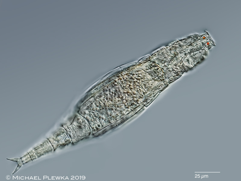
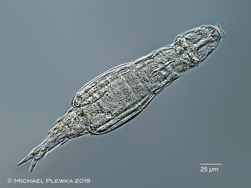
| Adineta cf oculata, dorsoventral view, creeping specimen. This is one of the two Adineta-species with eyespots. |
 |
| Adineta cf oculata, another creeping specimen from the same sample (dorsoventral view). While the eyespots in the first image are located in the rostrum (see the images below as further evidence) the eyspots in this 2nd image seem to be located "frontal"; their position in this image corresponds exactly to the position in the drawing of Adineta ricciae in the paper of Segers & Shiel (2005), which is obviously a misinterpretation of the microscopic view. |
| |
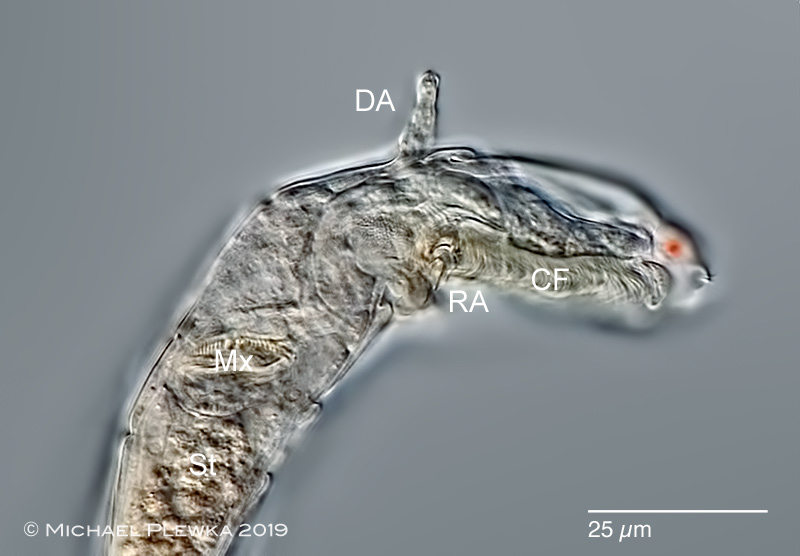 |
| Adineta cf oculata, anterior part, lateral view. This image shows that -in contrast to Adineta ricciae- the red eyespot (which is out of focus) is located in the rostrum. CF: ciliary field; DA: dorsal antenna; Mx: Mastax; RA: rake apparatus; St: stomach. |
| |
|
|
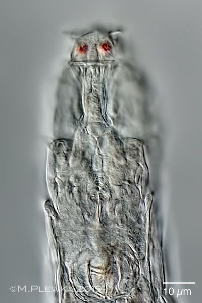
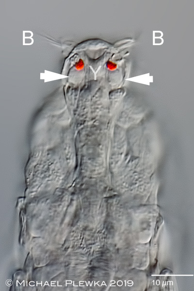
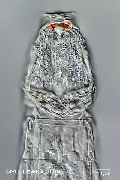
| Adineta cf oculata, 4 aspects of the head; focus plane from dorsal to ventral. Upper left: focus plane on the rostrum / first pseudosegment. Upper right: focus plane on the red eyespots (without lens). The arrowheads point to some structures that might be cells connected to the eyespots. Y: marks a Y-shaped structure (??nerve cell??) that yields at at the anterior ends in the bristles (B). Lower left: focus plane on the rostrum lamella and the hypodermis cells. The arrows point to some sensory cilia which are very short. The arrowheads point to (part of) the "stings" (see image below) which are connected to the rake apparatus. Lower right image: focus plane on the ventral ciliary field and rake apparatus. The U-hooks of the rake apparatus are folded backwards in this image. |
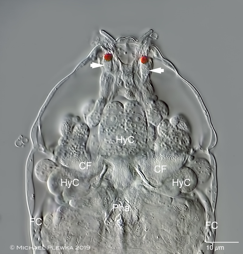 |
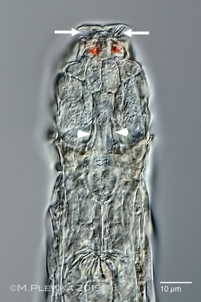
Adineta cf oculata, head of another specimen, slightly compressed by coverslide. Again the arrowheads point to some structures that might be cells which are connected to the red eyespots. Because of the compression or lack of oxygen the bristles in rostrum lamella have desintegrated into bubbles. HyC: hypodermis cells (in some of them the nucleus can be seen). CF: part of the ciliary field in the area of the rake apparatus. FC: flame cells. Pha: pharynx. |
| |
|
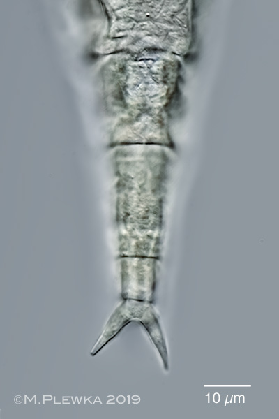
| Adineta cf oculata, two aspects of the foot. Left: focus plane on the spurs. Right: focus plane on the 3 toes (optical transect). |
| |
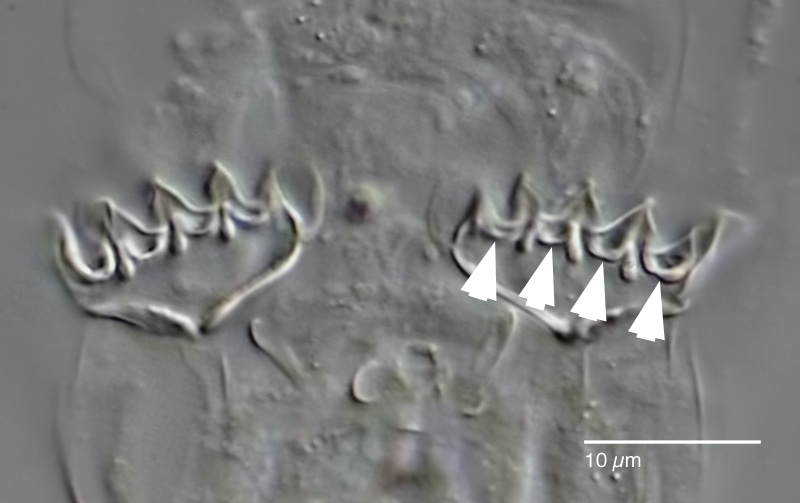 |
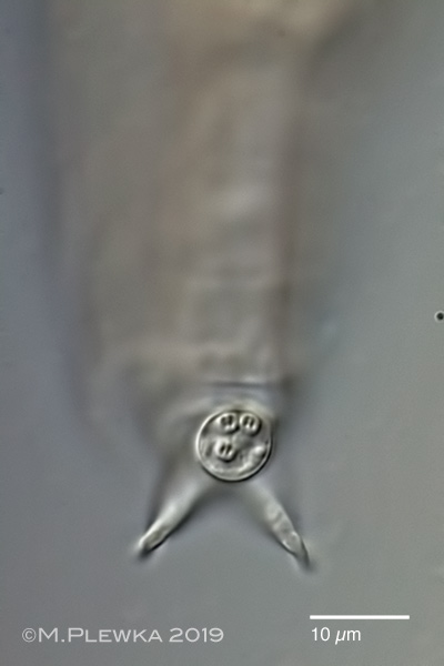
Adineta cf oculata, rake apparatus of a specimen treated with SDS, ventral view. The arrowheads point to the 4 U-hooks of the left rake. |
|
|
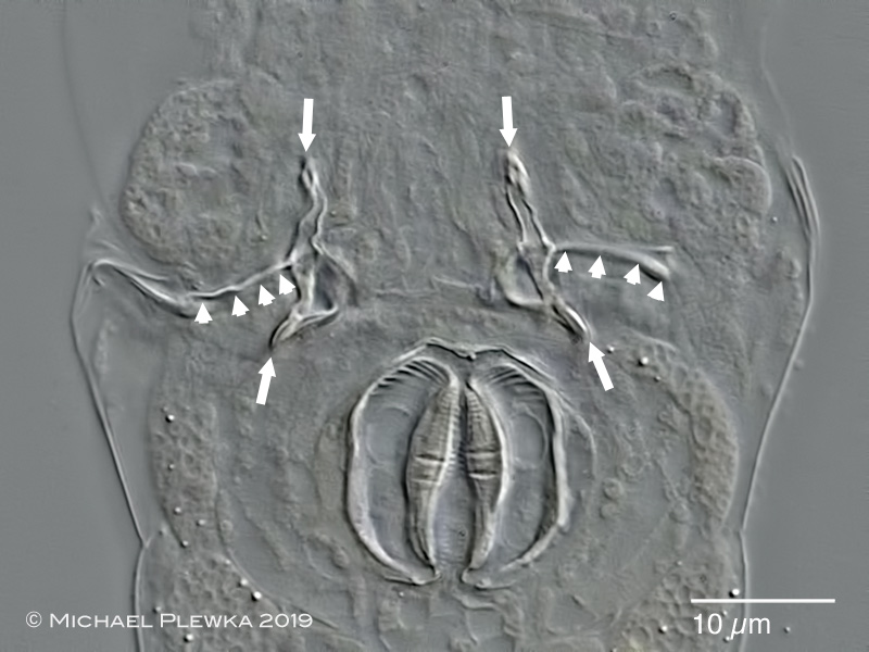 |
Adineta cf oculata, another specimen, treated with SDS and slightly compressed. The arrowheads point to the anterior rim of the basal plate of the rake apparatus (the denticles/ U-hooks are out of focus here). The arrows mark the "sting", a cuticularized structure of unknown function in the head. Also visible are the trophi. |
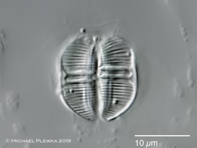 |
| Adineta cf oculata, trophi. |
| |
| |
| |
| Location: artificial culture material of unknown origin. |
| Habitat: aquatic sample. Sample courtesy of Dr. Renate Radek / FU Berlin and Dr. Sebastian Hess/ University Cologne |
| Date: 15.12.2019 |